名前:七條 敬文【国立がん研究センター中央病院 血液腫瘍科】
発表日時:2018年6月15日
発表形式:Poster
Title:
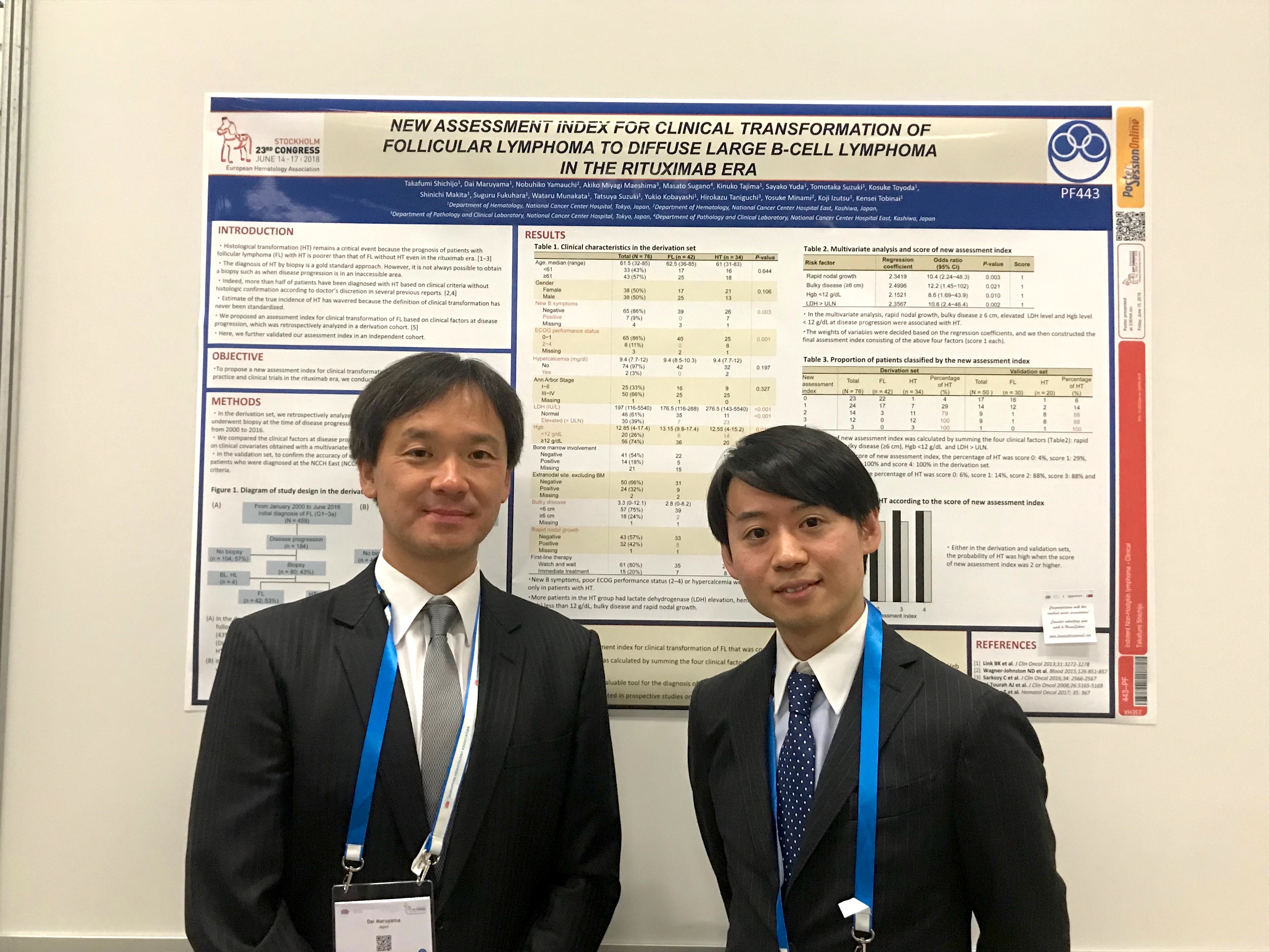
New assessment index of clinical transformation from follicular lymphoma (FL) to diffuse large B-cell lymphoma (DLBCL) in the rituximab era
Authors:
Takafumi Shichijo1, Dai Maruyama1, Nobuhiko Yamauchi2, Akiko Miyagi Maeshima3, Masato Sugano4, Kinuko Tajima1, Sayako Yuda1, Tomotaka Suzuki1, Kosuke Toyoda1, Shinichi Makita1, Suguru Fukuhara1, Wataru Munakata1, Tatsuya Suzuki1, Yukio Kobayashi1, Hirokazu Taniguchi3, Yosuke Minami2, Koji Izutsu1, Kensei Tobinai1
Affiliations:
1 Department of Hematology, National Cancer Center Hospital, Tokyo, Japan
2 Department of Hematology, National Cancer Center Hospital East, Kashiwa, Japan
3 Department of Pathology and Clinical Laboratory, National Cancer Center Hospital, Tokyo, Japan
4 Department of Pathology and Clinical Laboratory, National Cancer Center Hospital East, Kashiwa, Japan
Abstract:
Background: Histological transformation (HT) is still a critical event because the prognosis of FL patients (pts) with HT is poorer than that of FL pts without HT even in the rituximab era. Although the histological diagnosis of HT by biopsy is the gold standard, it is not always possible to obtain a biopsy specimen. Indeed, more than half of the pts have been diagnosed with HT based on clinical criteria without histological confirmation according to the doctor's discretion in several previous reports. Hence, estimates of the true incidence of HT have wavered because the definition of clinical transformation has never been standardized. Thus, we proposed an assessment index for clinical transformation, which was retrospectively analyzed in a derivation cohort [Hematol Oncol. 2017; 35: 367]. Here, we further validated our assessment index in an independent cohort.
Aims: To propose a new assessment index for clinical transformation of FL that is easy to use in both daily practice and clinical trials in the rituximab era, we conducted a multicenter retrospective analysis.
Methods: In the derivation set, we retrospectively analyzed pts who were initially diagnosed with FL and underwent biopsy at the time of disease progression at the National Cancer Center Hospital (NCCH) from 2000 to 2016. We compared the clinical factors at disease progression and constructed an assessment index based on clinical covariates obtained with a multivariate logistic regression model. In the validation set, to confirm the accuracy of our assessment index, we retrospectively analyzed pts who were diagnosed at the NCCH East (NCCHE) from 2003 to 2014 based on the same criteria.
Results: In the derivation set, 459 pts were diagnosed with FL (grade 1-3a) at the NCCH with a median follow-up duration of 9 (range: 0.7-16) years. Disease progression was observed in 184 pts, 80 (43%) of whom had histological documentation (FL in 42, HT with DLBCL in 34, HT other than DLBCL in 4). Finally, we identified 76 pts with biopsy-proven FL or HT with DLBCL as subjects for the derivation analysis. HT occurred at a median of 5.5 (range: 0.2-16) years from the initial FL diagnosis and the 5-year overall survival rate from HT was 62%. In the multivariate analysis, rapid nodal growth, bulky disease ≥6 cm, elevated serum lactate dehydrogenase (LDH) level and hemoglobin level (Hgb) <12 g/dl at disease progression were associated with HT. The weights of variables were decided based on the regression coefficients, and we then constructed the final assessment index consisting of the above four factors (score 1 each). The percentage of HT was score 0: 4%, score 1: 29%, score 2: 79%, score 3: 100% and score 4: 100%. In the validation set, 243 pts were diagnosed with FL (grade 1-3a) at NCCHE. Disease progression was observed in 95 pts, 50 (53%) of whom had histological documentation (FL in 30, HT with DLBCL in 20). Finally, our assessment index was applied to 50 pts with biopsy-proven FL or HT with DLBCL, and the percentage of HT was score 0: 6%, score 1: 14%, score 2: 88%, score 3: 88% and score 4: 100%. The probability of HT was high when the score was 2 or higher.
Conclusions: We developed a new assessment index for clinical transformation of FL that was confirmed by validation analysis. It is likely to be a simple and valuable tool for the diagnosis of HT, especially for pts from whom it is difficult to obtain a biopsy specimen. Further investigation is warranted in prospective studies on a large number of pts.
EHA2018参加レポート
この度は23rd EHA congressの参加にあたり,日本血液学会EHA Congress Travel Awardに採択いただき,誠にありがとうございました。
今年のEHA congressはストックホルムで開催され,世界各国から約12,000人近くの大勢の参加者で賑わいました。ストックホルムの中心部は海に面した中世の宮殿や建造物などの美しい景観が魅力的な都市でしたが,会場のあるAlvsjoは中央駅から電車で15分ほど離れた静閑な場所でした。しかし,会場内は活気に満ちており,どのセッションでも非常に活発な議論が見受けられました。EHA congressは教育セッションが多く,私のような若手にとっても非常に勉強になりました。私は,最新の臨床試験や,近年トピックである免疫療法,そして欧米で盛んなliquid biopsyのセッションなどを中心に聴講しました。特に欧米では,臨床試験と基礎的な解析を組み合わせた研究が多くされている印象であり,臨床試験と基礎的な分野のそれぞれを深く学び,コラボレートする必要性を感じました。
今回私は,"indolent lymphomaからaggressive lymphomaへ形質転換が疑われる患者が生検できない場合に,臨床的形質転換を定義する明確な規準がない"というclinical questionを基に,臨床的形質転換を定義する評価指標を提案するために臨床研究を行いました。具体的には,国立がん研究センター中央病院で初回診断および診療を受けた濾胞性リンパ腫患者459人の中で,病勢悪化時に生検を施行され,病理組織学的診断が確認された患者(濾胞性リンパ腫の再発,もしくはびまん性大細胞型B細胞リンパ腫への形質転換)を対象に,形質転換に関連する病勢増悪時の臨床因子を同定・スコア化し,評価指標を作成しました。引き続き,中央病院の患者コホートで作成した評価指標を国立がん研究センター東病院の患者コホートで検証した結果を含めて今回EHAで報告しました。Poster発表は1時間30分と決められた時間に会場を回り,それぞれの発表に興味を持った方と質疑応答を行うという形式でした。慣れない英語での議論でもあり,最初の30分程は時間が過ぎるのが遅いと感じましたが,その後はあっという間に時間が過ぎました。研究内容を海外の方から評価していただけるとやはり嬉しいもので,研究の励みとなり,より一層頑張ろうという気持ちになりました。また,印象に残った点としては,海外の若手研究者は物怖じせずに積極的に質問・議論をしており,日本の若手研究者も積極的な姿勢が必要だと肌で感じ,今後に向けて良い経験となりました。
最後になりますが,本研究をEHA Congress Travel Awardとして採択いただき,このような機会を与えて下さった日本血液学会の関係各位に心より感謝申し上げます。また,本研究の指導医である国立がん研究センター中央病院の丸山大先生,東病院患者コホートのデータ収集をお願いした国立がん研究センター東病院の山内寛彦先生,また,飛内賢正先生,伊豆津宏二先生をはじめとして本研究に関わっていただいた全ての皆様にこの場を借りて厚く御礼申し上げます。


