
名前:谷崎 祐太【早稲田大学 教育・総合科学学術院 分子生理学教室】
発表日時:2016年6月10日~11日
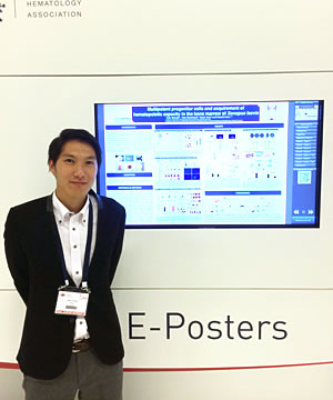
発表形式:e-poster
Title:
Multipotent progenitor cells and acquirement of hematopoietic capacity in the bone marrow of Xenopus laevis
Authors:
Yuta Tanizaki1, Yoko Mochizuki2, Takaki Aiso2 and Takashi Kato1, 2
Affiliations:
1Faculty of Education and Integrated Arts and Sciences, Waseda University, Tokyo, Japan;
2Integrative Bioscience and Biomedical Engineering, Graduate School of Advanced Science and Engineering, Waseda University, Tokyo, Japan;
Abstract:
Background
Hematopoietic stem cells (HSCs) are characterized by their capacity for long-term repopulation and multilineage differentiation upon cytokine stimulation. Thrombopoietin (TPO) is one of the key regulators of HSC maintenance and platelet production. Previously, we cloned Xenopus laevis (X. laevis) TPO (xlTPO) and demonstrated that differentiation and proliferation in thrombocyte progenitors and hematopoietic progenitors are regulated by xlTPO. Unlike that of mammals, bone marrow (BM) of X. laevis is filled with adipose cells, and blood cells are mainly produced in the liver. However, functional contribution of fatty marrow for hematopoiesis is unclear, and multipotent hematopoietic progenitor is not yet identified in X. laevis.
Aims
In this study, we attempted to identify multipotent hematopoietic cells and evaluate the ability of hematopoiesis in fatty marrow in X. laevis.
Methods
The suspension cells in X. laevis were cultured in semi-solid culture system in the presence of xlTPO. Laparotomy was performed in X. laevis under anesthesia with MS222. The left liver was resected, and cultured with xlTPO for 24 days. The cultured cells were labeled by PKH26 and autologously transplanted by intracardiac injection. After 30 days, the X. laevis were killed and their right liver was analyzed by FACS. PKH26 labelled cultured cells were also injected into the blastocoels of X. laevis embryos at stage 8, which have no immune system. After 3 days, the labeled cells were analyzed by fluorescence microscope. For low-temperature exposure, cage containing X. laevis were transferred to an incubator set at 5°C for 12 days. For micro-computerized tomography (mCT) analysis, X. laevis were restrained in polystyrene foam restrainers, and the femurs were imaged by three-dimensional X-ray CT scan.
Results
Hepatic colonies stimulated by xlTPO could be cultured for more than 3 months, during which the cell number reached 1 × 10⁶, indicating that the cells divided at least 20 times. These colonies expressed erythrocyte-, thrombocyte-, leukocyte-, and HSC-specific markers. Moreover, these cells differentiated to thrombocytes and leukocytes in the presence of splenic-conditioned media. Liver cells obtained by partial hepatic resection were cultured in the semi-solid culture system in presence of xlTPO, and cells labeled with PKH26 were autologously transplanted. After 30 days, PKH26-positive cells were detected in the sinusoids of liver and spleen. Flow cytometric analysis showed that the PKH26-positive cells displayed low forward (FSC) and side (SSC) scatter and had thin-layered cytoplasm and round nuclei, which are typical features of mammalian HSCs. These results indicated that xlTPO regulates proliferation of hematopoietic progenitors, which can be engrafted and differentiated to multiple lineages. To enrich multipotent hematopoietic cells, we generated anti-xlMPl monoclonal antibodies and showed that anti-thrombocyte antibody (T12)-/xlMpl+/FSClow population was enriched in high nuclear/cytoplasm ratio-hematopoietic progenitors, and the ratio of these cells to all hepatic cells was 0.28%. Surprisingly, T12-/xlMpl+/FSClow cells were identified in the liver, spleen, and BM in normal state. Although BM in X. laevis comprises mostly of adipocytes, large colonies induced by xlTPO were identified in the BM. The protein expression patterns in these cells overlapped with those in colonies derived from the liver, demonstrating that multipotent hematopoietic progenitors were localized in the BM. To explore the hematopoietic capacity in the BM, X. laevis were exposed to 5°C, which led to pancytopenia. After exposure to low temperature for 12 days, the numbers of erythrocyte and multipotent progenitors were increased. mCT revealed that femoral bone density was higher than that before exposure to 5°C, indicating change in the BM microenvironment.
Conclusion
In conclusion, multipotent hematopoietic progenitors in X. laevis are enriched in T12-/Mpl+/FSClow cells and these cells are mainly localized in the liver and BM. These findings suggested that T12-/Mpl+/FSClow cells in BM is dormant, and changes in the BM microenvironment by low-temperature stimulation induced proliferation of hematopoietic progenitors.
EHA2016参加レポート
この度は21st Congress of EHA参加にあたり,日本血液学会EHA travel awardに採択して頂きありがとうございました。学会会場であるコペンハーゲンは小さくまとまった街で生活しやすい印象を受けました。6月のコペンハーゲンは5時から22時まで明るく,学会が終わってからも街を散策することができた為,童話作家であるアンデルセンの世界観に満ちた街の空気を感じられました。これまでに参加したASH,ISEHと比較すると,その規模は中間に位置するような学会であり,ヨーロッパからの参加者が大半を占めておりました。EHAは若手をバックアップする姿勢を前面に押し出していることもあり,教育講演,ワークショップセッションが多く,私のような者にとって特に有意義な会となりました。夜に開催される若手の集まる会では日本語にも訳されているアット・ザ・ベンチを配り,学会会場では若手の質問者に対して菓子を配るなどユニークな取り組みが目立ちました。
多くの臨床医が参加する中で私の発表は数少ない非哺乳動物モデルを対象とした演題でした。私が研究対象にしているアフリカツメガエルは哺乳動物と比較すると抗体等利用できる道具が少なく解析には不利な点もありますが,環境変化に強く造血の環境応答を解析する上では最適なモデルの一つです。アフリカツメガエルの血球は主に肝臓で造られており,骨髄は健常な成人とは異なり脂肪髄を呈します。私は未同定であったアフリカツメガエル造血幹・前駆細胞を同定して,その細胞を指標として低温刺激における造血応答を解析しました。その結果,脂肪髄内に造血幹・前駆細胞が局在し,低温刺激が入ると骨髄で造血スイッチが入り,骨髄造血を開始することが明らかとなりました。本事象が再生不良性貧血における造血機能の獲得に関するモデルの一つになるのではないかと期待して今回の学会に臨みました。近年造血ニッチに関する報告が多く出てきていますが,今回の学会ではストレス環境下や各病態モデルにおける造血ニッチについて多くの議論が交わされていました。私が対象としている脂肪髄と造血に関する報告もその一部であり,早く論文にする必要性を感じました。今回の学会では一人での参加であることに加えて,e-poster発表の為,自身で議論する人を見つけなければならなく心配しておりましたが,その心配も杞憂に終わり,ポスター会場での議論は活発であり,その流れで自身の発表についても深く議論ができました。但し,あくまで主体的に動かないとその機会を得られないのが大変なところでした。議論の中では再生不良性貧血における免疫療法の観点から,低温時における,免疫応答の関与に関する質問が多く出ました。我々のような医学部のない大学での研究では,臨床の観点から見た議論がなかなか出来ないので,生の臨床医の声を聴くいい機会となりました。
今回,EHA travel awardとして採択して頂いた御蔭で,同年代の研究者と交流,議論ができて,今後の研究生活を送る上で非常に良い刺激を受けることが出来ました。今回の素晴らしい機会を与えて下さいました日本血液学会国際委員会の諸先生方,事務局の皆様,平素よりご指導頂いている早稲田大学・加藤尚志教授,研究を支えて下さいました皆様に心より感謝申し上げます。