

名前:濱中 有理【大阪大学大学院医学系研究科 血液・腫瘍内科】
発表日時:2016年6月11日
発表形式:ポスター
Title:
Anamorsin is essential for B-cell terminal differentiation
Authors:
Yuri Hamanaka, Hirohiko Shibayama, Akira Tanimura, Takafumi Yokota, Sachiko Ezoe, Michiko Ichii, Tomohiko Ishibashi, Yukiko Doi, Yasuhiro Nagate, Kenji Oritani, Yuzuru Kanakura
Affiliations:
Department of Hematology and Oncology, Osaka University Graduate School of Medicine
Abstract:
Background
Anamorsin (AM, also called CIAPIN1) is a cell death-defying factor, which we have cloned as a molecule that conferred resistance to apoptosis induced by growth factor deprivation. AM-deficient mice die during late gestation, and AM KO embryos are anemic and very small compared to wild-type embryos (J Exp Med 2004; 199: 581-592), suggesting that AM is essential for embryo growth and hematopoiesis. The embryonic lethality of AM KO mice impedes detailed analysis of the roles of AM. AM is important in Fe-S cluster protein biogenesis (BBRC 2011; 408: 329-333), which is indispensable in many biological processes, suggesting that AM may be required for critical biological pathways.
Aims
To overcome the embryonic lethality of AM KO, here we generated AM conditional KO (AMflox/flox) mice. The Cre-loxP system enabled cell type-specific genetic modification. Since we previously reported AM overexpression in ~30% of B-cell-type malignant lymphoma, here we focused on the role of AM in B-cell development.
Methods
We generated and analyzed CD19-Cre/AMflox/flox mice, with AM deleted specifically in CD19+ B cells (from Pro-B cells to mature peripheral B cells in B-cell development). B-cell development progresses through the pro-B cell, pre-B cell and immature B cell stages in BM and then exit from BM to spleen, where they pass through transitional stages; transitional type 1 (T1) and transitional type 2 (T2), and mature to either follicular B cells or marginal zone B cells.
Results
Compared with control CD19-Cre mice, CD19-Cre/AMflox/flox mice exhibited fewer B220+ B cells in the spleen (40.8±2.6% vs. 56.8±3.8%, P<0.001), peripheral blood (23.2±2.5% vs. 51.0±4.4%, P<0.001), and LN (8±2.3% vs. 31.7±1.0%, P<0.001). To further examine how AM deficiency impacted B-cell differentiation, BM and spleen cells were categorized into B-cell subsets -from hematopoietic stem cells to mature follicular and marginal zone B cells- based on marker expression patterns using flow cytometry. Compared to control mice, CD19-Cre/AMflox/flox mice showed significantly fewer follicular type I (FO-I) cells (B220+, AA4.1-, sIgMlow, CD23+, sIgD+), which are the most matured follicular B cells in the spleen (7.9±0.1% vs. 15.1±1.0%, P<0.01). Immunohistochemistry using monoclonal anti-B220 antibody in formalin-fixed paraffin-embedded spleen sections showed smaller B-cell follicles in CD19-Cre/AMflox/flox mice. Since AM was originally isolated as an anti-apoptotic molecule, spleen cells were treated with FITC-conjugated annexin V to determine the percentage of apoptotic cells, revealing greater apoptotic cell accumulation in CD19-Cre/AMflox/flox mice compared to controls (30.0±4.4% vs. 19.8±1.3%, P<0.05). This suggested that follicular B cell development was blocked by increased apoptosis in the CD19-Cre/AMflox/flox mice. Furthermore, CD19-Cre/AMflox/flox mice lacked splenic CD19+B220-/low cells, which display a plasmablast phenotype.
Conclusion
Although AM expression was consistent throughout the differentiation stages of CD19+ B cells, AM deficiency predominantly affected terminal differentiation of B cells, especially from T2 to FO-I. Immature B cells mature through transient T2 B cells into follicular or marginal zone B cells, and their follicular versus marginal zone fate partly depends on the strength of BCR signaling. Reduced numbers of FO-I B cells and CD19+B220-/low cells in the spleen are also reported in mice with defects in Bruton’s tyrosine kinase (BTK), which is activated in downstream of BCR signaling. The phenotypic similarity between Btk-deficient mice and our CD19-Cre/AMflox/flox mice suggests that AM may affect B-cell development through the BCR-BTK pathway. Overall, our present results show that AM plays essential roles in B-cell terminal differentiation, suggesting the potential involvement of some Fe-S cluster proteins in B-cell terminal differentiation. Moreover, our novel AMflox/flox mice will enable further investigation of the functions of AM and the related Fe-S cluster proteins in various organs.
EHA2016参加レポート
この度は、21th Congress of European Hematology Association(EHA)で発表するにあたって、Travel Awardに採択していただき、誠にありがとうございました。学会はデンマークの首都、コペンハーゲンにて2016年6月9日から12日まで開催されました。コペンハーゲンは、300年以上前に建設された中世の建物が今も残り、運河沿いにはカラフルな家が並ぶ美しい街並みで、まさにおとぎの国という雰囲気でした。会場となったベラ・センターは、市街地からは少し離れた郊外にありましたが、地下鉄の駅前にあり街の中心部から容易にアクセスできました。会場は広く、同時にいくつものセッションが行われ、ヨーロッパはもちろん、アメリカやオーストラリア、アジアなどから多くの参加者が集まる活気溢れる雰囲気に、海外学会の経験が少ない私は、最初は圧倒されましたが、活発な意見交換や同年代の方が堂々と発表している姿から刺激を受け、日本に帰国してから、心新たに頑張ろうという気持ちになりました。
現在、臨床業務に携わっていることから、臨床に関連しているセッションを中心に参加しました。再発/難治性B前駆細胞型ALLに対するブリナツモマブ、多発性骨髄腫に対するダラツムマブなどの新規薬剤に関する最新の知見を得ることができました。また普段、話を聴く機会の少ない、肝移植における制御性T細胞や腎移植における同種Mesenchymal Stromal Cellsを用いた免疫寛容の誘導など、臓器移植におけるトレランスの話は興味深く拝聴しました。
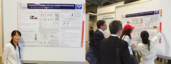
また、今回、私は、当科で10年程前に抗アポトーシス分子として同定したアナモルシンのBリンパ球分化における役割について発表いたしました。アナモルシンのノックアウトマウスは胎生後期に致死となり、胎仔は小さく貧血様であることから、アナモルシンは胎生期の造血において重要な役割をしていると考えられていますが、成獣での造血における役割は不明でした。そこで、アナモルシンのコンディショナルノックアウトマウスを初めて作製し、CD19-Creマウスと交配してB細胞特異的アナモルシン欠損マウスを作製しました。解析の結果、脾臓におけるBリンパ球分化の最終段階である、Follicular type I細胞が減少し、またそれ以降の末梢血やリンパ節における成熟Bリンパ球が減少しており、アナモルシンがBリンパ球の最終分化において重要な役割をしていることを見出しました。今後はこのコンディショナルノックアウトマウスを使用し、種々の臓器や細胞におけるアナモルシンの機能解析を進めていこうと考えています。アナモルシンはまだまだ認知度が低い分子であり、どれくらいの人に興味を持っていただけるか不安でしたが、想像以上に多くの人が話を聴いてくださり、緊張もしましたが、とても嬉しい経験でした。
最後になりますが、日頃よりご指導を頂いております柴山浩彦先生、金倉譲教授を始め医局の先生方、貴重な機会を与えてくださった日本血液学会国際委員会の先生方、事務局の皆様に心より感謝申し上げます。