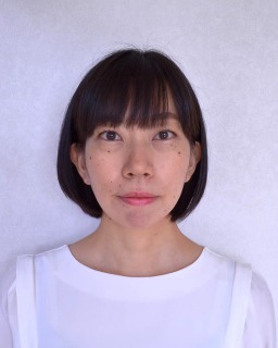
名前:須摩 桜子【筑波大学 人間総合科学研究科 疾患制御医学専攻】
発表形式:e-Poster
Title:
Single-cell RNA sequencing reveals tumor cell heterogeneity and comprehensive immune profile of T follicular helper cell lymphoma
Authors:
Sakurako Suma1, Manabu Fujisawa2, Yoshiaki Abe1, Yasuhito Suehara2, 3, Daisuke Kaji4, Takeshi Sugio5, Koji Kato6, Koichi Akashi6, Kosei Matsue7, Naoya Nakamura8, Ayako Suzuki9, Yutaka Suzuki9, Shigeru Chiba2, 3, and Mamiko Sakata-Yanagimoto2, 3, 10
Affiliations:
1. Department of Hematology, Graduate School of Comprehensive Human Sciences, University of Tsukuba, Tsukuba, Japan.
2. Department of Hematology, Faculty of Medicine, University of Tsukuba, Tsukuba, Japan.
3. Department of Hematology, University of Tsukuba Hospital, Tsukuba, Japan.
4. Department of Hematology, Toranomon Hospital, Tokyo, Japan.
5. Department of Medicine, Division of Oncology, Stanford University, Stanford, United States.
6. Department of Medicine and Biosystemic Science, Kyushu University Graduate School of Medical Science, Fukuoka, Japan.
7. Division of Hematology/Oncology, Department of Internal Medicine, Kameda Medical Center, Kamogawa, Japan.
8. Department of Pathology, Tokai University School of Medicine, Isehara, Japan.
9. Department of Computational Biology and Medical Sciences, The University of Tokyo, Tokyo, Japan.
10. Division of Advanced Hemato-Oncology, Transborder Medical Research Center, University of Tsukuba, Tsukuba, Japan.
Abstract:
Background: Angioimmunoblastic T-cell lymphoma (AITL) and other nodal lymphomas of T follicular helper (TFH) cell origin (TFH lymphoma) are associated with immune dysregulation. However, comprehensive immune profiles of TFH lymphoma have not been fully investigated. The RHOA G17V mutation (G17V) is found in 60-70% of TFH lymphoma and considered to gain a novel function through binding to VAV1 protein. Nevertheless, the prevalence of homozygous (Homo) and heterozygous (Hetero) G17V mutations and the difference in their oncogenicity due to its zygosity remain unclear.
Aims: We performed single-cell RNA sequencing (scRNA-seq) for immune and tumor cells of TFH lymphoma to determine (1) the specific immune profiles, (2) the characteristics of tumor cells and (3) the mechanisms of clonal evolution from G17V Hetero to Homo.
Methods: We collected 29 PB (12 at initial diagnosis [ID], 17 at relapsed/refractory diagnosis [RR]) and 10 LN (6 ID, 4 RR) from 25 patients with TFH lymphoma, 7 homeostatic LN (HLN) from patients with solid cancers. scRNA-seq and single-cell T cell receptor (TCR) sequencing were performed by the Chromium system (10x Genomics) from 16 PB and 9 LN of TFH lymphoma patients and 7 HLN. Publicly available scRNA-seq data from 5 healthy donors (HD) were also integrated as controls. scRNA-seq data analysis was performed by Seurat, Scater and Batchelor for quality control, integration, and clustering; SingleR for annotation; Monocle3 and Slingshot for trajectory analysis; Immunarch for TCR repertoire analysis, and gene set variation analysis (GSVA) for pathway analysis. VarTrix pipeline was used to detect mutation distribution in scRNA-seq data.
Results: In total, 98,891 cells from PB and 68,738 cells from LN were analyzed. Unsupervised clustering identified 19 T/NK-, 2 B-, and 9 myeloid-cell clusters in PB (Figure 1). Tumor cells were identified by the expanded TCR clonality and the TFH-like gene expression signatures. (1) Subsequent clustering and GSVA of each cell type identified dysfunctional CD8 T cells (Dys CD8) in PB and LN of patients, some of which had enriched the proliferation signature (Pro CD8) (Figure 2). In some samples, Dys/Pro CD8 shared their TCRs between PB and LN, suggested that they have the same cellular origin. The proportion of Pro CD8 in LN correlated with that of TREG in PB; moreover, TREG effector function was activated in both PB and LN of patients compared to HD and HLN, respectively. By sub-clustering of myeloid subset, C1QA/B/C (C1Q) + macrophages and LAMP3+PD-L1+ dendritic cells (DCs) increased in LN of RR patients, whereas C1Q+ monocytes increased and CLEC9A+ conventional DCs and plasmacytoid DCs decreased in PB. (2) Differential expression gene (DEG) analysis identified five tumor-specific gene candidates and the cell surface protein PLS3 was validated by flow cytometry (Figure 3). (3) The G17V RHOA-mutated cells were detected in 6 PB and 3 LN. The G17V Hetero- versus Homo-mutated cells were 657 vs 59 in PB, while 1529 vs 288 in LN. DEG and GSVA in LN identified that the regulatory signature was significantly activated in Homo, whereas the proliferation signature was up-regulated in Hetero (Figure 4).
Summary/Conclusion: These results clarified the comprehensive immune structures including novel tumor-specific markers in TFH lymphoma. PLS3, one of the new tumor markers, could be detected by flow cytometry, a viable method in clinical practice. In the clonal evolution process from the G17V Hetero to Homo, the proliferative signaling switch to the immune escape signaling.
EHA2022参加レポート
この度はthe European Hematology Association (EHA) 2022 Hybrid Congressの参加にあたり、JSH Travel Award for EHA Congressに採択いただきまして、誠にありがとうございました。今年のEHA Congressは、現地とオンラインでのハイブリッド開催となり、私はオンラインから参加させていただきました。対面でのディスカッションや会場での臨場感を感じることができなかったのは残念でしたが、ライブ配信およびオンデマンド配信により、日本にいながらにして様々なプログラムを視聴することができ、大変有意義な時間となりました。
本学会において私は、血管免疫芽球性T細胞リンパ腫(AITL)をはじめとするT濾胞ヘルパー細胞起源のリンパ腫(TFHリンパ腫)のシングルセルRNAシークエンス解析について報告させていただきました。TFHリンパ腫、特にAITLの腫瘍組織においては、腫瘍細胞の周囲に反応性リンパ球や組織球、好酸球、形質細胞など種々の免疫細胞が浸潤し、腫瘍の発症・進展に大きく関わっていると考えられていますが、シングルセルレベルでの免疫プロファイルはまだ完全には解明されていません。また、我々の研究室ではこれまでに、TFHリンパ腫の60-70%でみられる疾患特異的遺伝子変異であるRHOA G17V変異を同定し、G17V変異体がVAV1蛋白と特異的に結合することでT細胞受容体(TCR)シグナルを増強し、リンパ腫形成に寄与していることを報告しております。今回我々は、TFHリンパ腫患者のリンパ節と末梢血のシングルセル解析を通して、TFHリンパ腫の包括的免疫プロファイルと腫瘍細胞の特徴を明らかにしました。TFHリンパ腫は予後不良な疾患であり、未だに標準治療が定まっていません。今回の報告が、今後の治療戦略改善の一助になればと考えております。
EHAの参加は今回が初めてでしたが、基礎・臨床研究ともに充実した大変内容の濃い学会でした。何より、私にとっては基礎研究を始めてから初の本格的な国際学会への参加であり、国際的に活躍されている先生方の研究へのアイデアや手法、解析技術などを知ることができ、非常に有意義な時間を過ごすことができました。T細胞リンパ腫に関する発表はB細胞リンパ腫に比して多くはありませんでしたが、自分の研究と関連した内容について、世界で行われている研究を垣間見ることができ、大変刺激になりました。
最後になりましたが、今回このような貴重な機会を与えていただきました日本血液学会国際委員会の先生方、日本血液学会事務局の皆様に心より感謝いたします。また、日頃ご指導いただいている千葉滋教授、坂田麻実子教授をはじめ、お世話になっている先生方、研究にご協力いただいている各施設の先生方ならびにスタッフの方々にこの場をお借りして深く御礼申し上げます。


