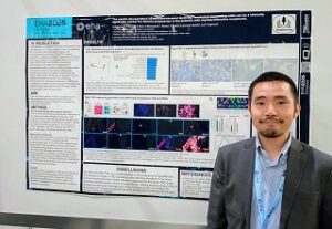
30th EHA Congress Travel Award 受賞レポート 永春 圭規
名前:永春 圭規【三重大学血液・腫瘍内科/ルンド大学分子血液学】
発表形式:Poster
Title:
The spatial dysregulation of microenvironment involving Tenascin-C expressing cell can be a clinically applicable marker for fibrosis progression in the patients with myeloproliferative neoplasms
Authors:
K Nagaharu1,2 , Y Kojima3 , E Ohya2 , K Maruayama4 , I Tawara2 , K Ohishi2 , K Imanaka-Yoshida4 , and Y Sugimoto2
Affiliations:
1. Lund University, Laboratory Medicine, Lund, Sweden
2. Hematology and Oncology, Mie University Graduate School of Medicine, Tsu, Japan
3. National Cancer Center Research Institute, Laboratory of Computational Life Science, Tokyo, Japan
4. Pathology and Matrix Biology, Mie University, Tsu, Japan
5. Transfusion and Cell Therapy, Mie University Hospital, Tsu, Japan
Abstract:
Background: Philadelphia chromosome-negative myeloproliferative neoplasms (MPNs) are a group of intractable hematological malignancies characterized by aberrant JAK-STAT pathway activation, ultimately leading to bone marrow fibrosis. Previous studies have implicated pathways such as JAK-STAT, NFκB, and SMAD3 in promoting inflammation and altering the expression of extracellular matrix proteins (ECMs) in bone marrow microenvironment during fibrosis. However, clinically applicable biomarkers to assess these microenvironmental changes of non-hematopoietic cells remain elusive.
Aims: This study aims to identify clinically applicable markers for evaluating the bone marrow microenvironment in MPN patients by focusing on ECMs as a potential early indicator of myelofibrosis.
Methods: We performed simultaneous staining of 28 molecules in normal (n=2), CALR-mutated Essential thrombocytosis (ET, n=2) and CALR-mutated Myelo-fibrosis (MF, n=2) using Akoya Phenocycler fusion. The extracted fluorescent values and coordinates of more than 500,000 cells were further analyzed by Scanpy (python). In parallel, we utilized the ELISA to validate the serum levels of highly probable clinical molecules. Written informed consent was obtained by all participants, and our study was approved by Mie University ethical committee.
Results: First, we challenged to identify the possible markers in MPNs patients during progression from human bone marrow single cell RNA seq repository data. Our in silico analysis led to identifying the increase of several ECMs in CD45- cells at ET stage and declining during MF progression. We hypothesized these dynamics could serve as clinical predictors for myelofibrosis. In line with previous reports, nestin+ CD45- cells are dynamically changed along with the disease progression. To validate clinical utility of these molecules, we proceeded to the multicolor-immunofluorescent staining of bone marrow specimen obtained from patient’s samples at the diagnosis. We employed the simultaneous multicolor staining of 28 antigens to reveal the characteristics of TNC expressing cells. Clustering using 28 antigens showed a large part of CD45- cells are classified into LEPR+ mesenchymal stem-like cells, endothelial cells, TNC expressing CD45- cells and others. The comparison between disease state, TNC expressing cells showed a 5.5 and 2.5-fold increase in ET and myelofibrosis samples, respectively, compared to healthy donors. Spatial analysis using several molecules and coordinates of specimen revealed low proximity score between TNC expressing cells and other CD45- cells as the MPN progress. These results indicate that microenvironments regarding TNC expressing cells differ during myelofibrosis. We evaluated clinical utility of serum TNC levels in patients with ET and MF (n=55), and found that serum TNC was significantly lower in the MF stage (mean 25.7 ng/mL) compared to the ET stage (mean 91.3 ng/mL, p-value < 0.001).
Summary/Conclusion: This study identifies TNC as a clinical indicator in the progression of myelofibrosis, with dynamic changes in TNC-expressing cells reflecting alterations in the bone marrow microenvironment. Besides, our data suggested that the serum TNC levels could be a promising early marker for myelofibrosis, offering potential clinical utility in disease monitoring and management. These findings provide a foundation for developing non-invasive diagnostic tools and advancing the understanding of bone marrow dynamics in MPNs.
EHA2025参加レポート
この度はJSH-EHA Travel Grant Programに採用いただき、2025年のEHA annual congressに参加してまいりました。同会は6月のイタリア・ミラノにおいて開催され、摂氏35-37度の猛暑の中、EHA会場は外気に負けず熱気を含んだものでした。今回、私は「骨髄増殖性腫瘍の臨床応用可能なbiomarker」に関する発表機会を得ることができました。本研究は、私自身抱いてきた日常診療のジレンマを解決するため始めたプロジェクトで、参画いただいた先生方との日々の発見をもとに行ってきた研究です。気が付けば、診療科の枠を超えた共同研究に至り、今後のさらに発展させることを目指しています。今回の発表は諸先生方のご指導と、科研費および先進ゲノム支援事業によるサポートによって、ここまで発展させることができました。いろいろな形でご指導いただきました先生方には感謝しています。
このような海外の大きな学会に参加させていただく機会は得難い経験であり、EHA学会会場では積極的にdiscussionへの参加を目指しました。最先端のBioinformatics分野の若手研究者や、造血幹細胞研究で著名なDr. Laurentiなど論文を通じてしか名の知らなかった先生とも直接のやり取りができたことはかけがえのない財産となりました。中でも印象に残ったこととして、私自身何度も引用させていただいたDr. Barbui Tiziano (本態性血小板血症のリスク予測であるIPSETの著者)と直接話ができた上、以前の自分の仕事を認知していただいていたことを受け、とても心揺さぶれる体験ができました。
EHAを通じて感じた本学会のエネルギーは大きく2つあり、1つ目は多くの発表がinternational studyに基づくものであること。欧州諸国は1国の規模ではUSA、Asiaより少なくなることが多い分、臨床・基礎両面での国としての垣根を低くしたnetworkingが強い推進力となっていることを感じました。そして2つ目は若手の積極性です。若手の研究者のテーマセッションが多く、chairも若年の方が多いという点も印象を受けました。通常の発表だけではなく複数のdiscussion baseのセッションが開かれ、異分野であってもその双方向のやりとりは大変興味深いものでした。私はポスター発表でしたが、質問に来ていただいた研究者の中には類似した知見を示唆する内容と非常に鋭い意見もいただき、身の引き締まる思いを抱くとともに明日からの研究を、さらに有意義なものにすべくエネルギーを得ることができたと感じています。
最後に改めまして、EHA2025の参加を支援いただきました三重大学血液内科・修復再生病理学の先生方、このような機会を設けていただきましたJSH・EHA学会関係者の皆様に感謝いたします。